Ankle fracture: Difference between revisions
(sensitivity) |
|||
| (36 intermediate revisions by 7 users not shown) | |||
| Line 1: | Line 1: | ||
== | {{Adult top}} [[ankle fracture (peds)]] | ||
==Background== | |||
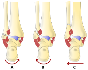
[[File:Weber Classification - latin.png|thumb|Danis–Weber classification of ankle fractures (Types A, B and C).]] | |||
==Clinical Features== | |||
*Examine for ecchymoses, abrasions, or swelling | *Examine for ecchymoses, abrasions, or swelling | ||
* | *Vascular and neurologic assessment | ||
** | **DP and PT pulses | ||
**4 sensation distributions: saphenous nerve (medial mal), superficial fib (lat mal), sural nerve (lateral 5th digit), deep fib (1st web space) | **4 sensation distributions: saphenous nerve (medial mal), superficial fib (lat mal), sural nerve (lateral 5th digit), deep fib (1st web space) | ||
*Note skin integrity and areas of tenderness or crepitus over ankle | *Note skin integrity and areas of tenderness or crepitus over ankle | ||
*Range joint passively and actively to evaluate for stability | *Range joint passively and actively to evaluate for stability | ||
*Examine | *Examine joints above and below the ankle | ||
*Perform anterior drawer test (positive exam suggests torn ATFL) | *Perform anterior drawer test (positive exam suggests torn ATFL) | ||
*'''Always palpate entire length of fibula to rule-out [[Maisonneuve Fracture]] (fibulotibialis ligament tear)''' | *'''Always palpate entire length of fibula to rule-out [[Maisonneuve Fracture]] (fibulotibialis ligament tear)''' | ||
| Line 13: | Line 17: | ||
*Palpate midfoot and base of 5th metatarsal for tenderness | *Palpate midfoot and base of 5th metatarsal for tenderness | ||
==Diagnosis== | ==Differential Diagnosis== | ||
*[[Ottawa Ankle Rules]] (sen 96-99% for excluding | {{Other ankle injuries DDX}} | ||
{{Distal leg fractures DDX}} | |||
{{Foot and toe fractures DDX}} | |||
==Evaluation== | |||
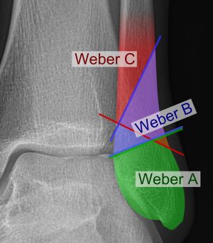
[[File:Danis–Weber classification on X-ray.jpg|thumb|Danis–Weber classification on X-ray.]] | |||
[[File:WeberARadiopediaOB.jpg|thumb|Weber A Oblique]] | |||
[[File:WeberBRadiopedOB.jpg|thumb|Weber B Oblique]] | |||
[[File:WeberBAPMedp.jpg|thumb|Weber B AP]] | |||
[[File:WeberCOBMedp.jpg|thumb|Weber C Oblique]] | |||
[[File:WeberCAPMedp.jpg|thumb|Weber C AP]] | |||
*[[Ottawa Ankle Rules]] (sen 96-99% for excluding fracture) | |||
*3 views: | *3 views: | ||
**AP | **AP: Best for isolated lateral and medial malleolar fractures | ||
**Oblique (mortise) | **Oblique (mortise) | ||
***Best for evaluating for unstable fracture or soft tissue injury | ***Best for evaluating for unstable fracture or soft tissue injury | ||
***At a point | ***At a point 1 cm proximal to tibial plafond space between tib/fib should be ≤6mm | ||
**Lateral | **Lateral: Best for posterior malleolar fractures | ||
*Consider proximal tib/fib films and talus fractures | |||
* | |||
==Classification (Danis-Weber System)== | ===Classification (Danis-Weber System)=== | ||
* | [[File:WeberclassRadioped.jpg|thumb|]] | ||
* | *System based on level of the fibular fracture and characterizes stability of fracture | ||
*Tibial plafond and the two malleoli is referred to as the ankle "mortise" (or talar mortise) | |||
===Type A=== | ====Type A==== | ||
* | *Fibula fracture below ankle joint/distal to plafond | ||
** | **Medial malleolus often fractured | ||
** | **Tibiofibular syndesmosis intact | ||
** | **Usually stable: occasionally requires ORIF | ||
===Type B=== | ====Type B==== | ||
* | *Fibula fracture at the level of the ankle joint/at the plafond | ||
** | **Can extend superiorly and laterally up fibula | ||
** | **Tibiofibular syndesmosis intact or only partially torn | ||
** | **No widening of the distal tibiofibular articulation | ||
** | **Medial malleolus may be fracture | ||
** | **Possible instability | ||
***Use gravity or weight bearing stress X-rays to determine stability <ref>Tips for Managing Weber B Ankle Fractures By Joseph Noack, MD; and Spencer Tomberg, MD. ACEP Now April 14, 2020 https://www.acepnow.com/article/tips-for-managing-weber-b-ankle-fractures/?singlepage=1</ref> | |||
===Type C=== | ====Type C==== | ||
* | *Fibula fracture above the level of the ankle joint/proximal to plafond | ||
** | **Tibiofibular syndesmosis disrupted with widening of the distal tibiofibular articulation | ||
** | **Medial malleolus fracture | ||
** | **Unstable: requires ORIF | ||
==Management & Disposition== | |||
{{General Fracture Management}} | |||
== | ===General Ankle Fracture=== | ||
*Determined by stability of fracture: | |||
**Stable, nondisplaced, isolated malleolar fracture: Splint or cast, early wt bearing, RICE | |||
**Unstable or displaced fracture: Requires ORIF, ortho consult, reduce and splint | |||
* | ===Isolated lateral malleolar fracture=== | ||
** | *If stable (see Weber classification) treat like severe [[Ankle Sprain]] | ||
** | *Signs of instability: | ||
**Displacement >3mm | |||
**Associated medial malleolus fracture | |||
**Signs of medial (deltoid) ligament disruption such as medial swelling, ecchymosis, or TTP | |||
**Widening of medial clear space (suggests deltoid ligament injury) | |||
===Isolated medial or posterior malleolar fracture=== | |||
*Must rule-out other injuries | |||
*If non-displaced, isolated: | |||
**[[Short-Leg Posterior Splint]] (ankle at 90<sup>o</sup>) | |||
**Non-weight bearing | |||
**Refer to Ortho in 5-7d | |||
== | ===Lateral malleolar fracture with deltoid injury OR bimalleolar OR trimalleolar fracture=== | ||
[[File: | [[File:Bimalleolar fracture legend.jpg|thumb|Bimalleolar fracture and right ankle dislocation on X-ray (anteroposterior). Both the end of the fibula (1) and the tibia (2) are broken and the malleolar fragments (arrow: medial malleolus, arrowhead: lateral malleolus) are displaced.]] | ||
*[[Short-Leg Posterior Splint]] (ankle at 90<sup>o</sup>) | |||
*Immediate reduction or ortho consult in ED | |||
[[ | |||
==See Also== | ==See Also== | ||
| Line 88: | Line 99: | ||
*[[Ankle Sprain]] | *[[Ankle Sprain]] | ||
*[[Ankle Fracture (Peds)]] | *[[Ankle Fracture (Peds)]] | ||
*[[ | *[[Ottawa Ankle Rules]] | ||
*[[Maisonneuve Fracture]] | *[[Maisonneuve Fracture]] | ||
*[[Pilon Fracture]] | *[[Pilon Fracture]] | ||
| Line 94: | Line 105: | ||
*[[Splinting]] | *[[Splinting]] | ||
== | ==External Links== | ||
*http://radiopaedia.org/articles/weber_ankle_fracture_classification (Images by Dr. Frank Gaillard; CC SA NC BY licence) | *http://radiopaedia.org/articles/weber_ankle_fracture_classification (Images by Dr. Frank Gaillard; CC SA NC BY licence) | ||
[[Category: | *Ottawa Ankle Rules - http://www.ncbi.nlm.nih.gov/pubmed?term=12595378 | ||
==References== | |||
<references/> | |||
[[Category:Orthopedics]] | |||
Latest revision as of 22:50, 5 March 2025
This page is for adult patients. For pediatric patients, see: ankle fracture (peds)
Background
Clinical Features
- Examine for ecchymoses, abrasions, or swelling
- Vascular and neurologic assessment
- DP and PT pulses
- 4 sensation distributions: saphenous nerve (medial mal), superficial fib (lat mal), sural nerve (lateral 5th digit), deep fib (1st web space)
- Note skin integrity and areas of tenderness or crepitus over ankle
- Range joint passively and actively to evaluate for stability
- Examine joints above and below the ankle
- Perform anterior drawer test (positive exam suggests torn ATFL)
- Always palpate entire length of fibula to rule-out Maisonneuve Fracture (fibulotibialis ligament tear)
- Perform a crossed-leg test to detect syndesmotic injury
- Evaluate integrity of Achilles tendon (Thompson test)
- Palpate midfoot and base of 5th metatarsal for tenderness
Differential Diagnosis
Other Ankle Injuries
Distal Leg Fracture Types
- Tibial plateau fracture
- Tibial shaft fracture
- Pilon fracture
- Maisonneuve fracture
- Tibia fracture (peds)
- Ankle fracture
- Foot and toe fractures
Foot and Toe Fracture Types
Hindfoot
Midfoot
Forefoot
Evaluation
- Ottawa Ankle Rules (sen 96-99% for excluding fracture)
- 3 views:
- AP: Best for isolated lateral and medial malleolar fractures
- Oblique (mortise)
- Best for evaluating for unstable fracture or soft tissue injury
- At a point 1 cm proximal to tibial plafond space between tib/fib should be ≤6mm
- Lateral: Best for posterior malleolar fractures
- Consider proximal tib/fib films and talus fractures
Classification (Danis-Weber System)
- System based on level of the fibular fracture and characterizes stability of fracture
- Tibial plafond and the two malleoli is referred to as the ankle "mortise" (or talar mortise)
Type A
- Fibula fracture below ankle joint/distal to plafond
- Medial malleolus often fractured
- Tibiofibular syndesmosis intact
- Usually stable: occasionally requires ORIF
Type B
- Fibula fracture at the level of the ankle joint/at the plafond
- Can extend superiorly and laterally up fibula
- Tibiofibular syndesmosis intact or only partially torn
- No widening of the distal tibiofibular articulation
- Medial malleolus may be fracture
- Possible instability
- Use gravity or weight bearing stress X-rays to determine stability [1]
Type C
- Fibula fracture above the level of the ankle joint/proximal to plafond
- Tibiofibular syndesmosis disrupted with widening of the distal tibiofibular articulation
- Medial malleolus fracture
- Unstable: requires ORIF
Management & Disposition
General Fracture Management
- Acute pain management
- Open fractures require immediate IV antibiotics and urgent surgical washout
- Neurovascular compromise from fracture requires emergent reduction and/or orthopedic intervention
- Consider risk for compartment syndrome
General Ankle Fracture
- Determined by stability of fracture:
- Stable, nondisplaced, isolated malleolar fracture: Splint or cast, early wt bearing, RICE
- Unstable or displaced fracture: Requires ORIF, ortho consult, reduce and splint
Isolated lateral malleolar fracture
- If stable (see Weber classification) treat like severe Ankle Sprain
- Signs of instability:
- Displacement >3mm
- Associated medial malleolus fracture
- Signs of medial (deltoid) ligament disruption such as medial swelling, ecchymosis, or TTP
- Widening of medial clear space (suggests deltoid ligament injury)
Isolated medial or posterior malleolar fracture
- Must rule-out other injuries
- If non-displaced, isolated:
- Short-Leg Posterior Splint (ankle at 90o)
- Non-weight bearing
- Refer to Ortho in 5-7d
Lateral malleolar fracture with deltoid injury OR bimalleolar OR trimalleolar fracture
- Short-Leg Posterior Splint (ankle at 90o)
- Immediate reduction or ortho consult in ED
See Also
- Ankle (Main)
- Ankle Sprain
- Ankle Fracture (Peds)
- Ottawa Ankle Rules
- Maisonneuve Fracture
- Pilon Fracture
- Fracture (Main)
- Splinting
External Links
- http://radiopaedia.org/articles/weber_ankle_fracture_classification (Images by Dr. Frank Gaillard; CC SA NC BY licence)
- Ottawa Ankle Rules - http://www.ncbi.nlm.nih.gov/pubmed?term=12595378
References
- ↑ Tips for Managing Weber B Ankle Fractures By Joseph Noack, MD; and Spencer Tomberg, MD. ACEP Now April 14, 2020 https://www.acepnow.com/article/tips-for-managing-weber-b-ankle-fractures/?singlepage=1


