Open-angle glaucoma: Difference between revisions
No edit summary |
|||
| Line 30: | Line 30: | ||
* Thinning or notching of disc rim | * Thinning or notching of disc rim | ||
* Progressive change of size/shape of cup | * Progressive change of size/shape of cup | ||
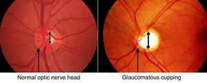
[[File:glaucoma-cupping-1024x414.jpg|thumb|Glaucoma cupping]] | |||
==== Visual Field testing ==== | ==== Visual Field testing ==== | ||
==== Intraocular pressure ==== | ==== Intraocular pressure ==== | ||
Revision as of 19:29, 29 November 2016
Open-angle Glaucoma
Open-angle glaucoma is an optic neuropathy characterized by an increase in intraocular pressure leading to damage to the optic nerve and irreversible vision loss. It most commonly presents with progressive peripheral vision loss, followed by central vision loss and is the second leading cause of irreversible blindness worldwide.
Pathophysiology
Not entirely clear but may be related to an increased intraocular pressure that leads to compression of the optic nerve at the site where it exits the eye. This causes a progressive decrease in the number of retinal ganglion cells.
Risk Factors
- Age. 4% prevalence in age >80
- Race- 3x higher in African Americans
- Family History. 2-3 fold increase for individuals with affected sibling or parent
- Hypertension
- Diabetes
- Other: Myopia, pseudoexfoliation, low diastolic perfusion pressure, CV disease, Hypothyroidism
Clinical Presentation
- Painless
- Cupping of the optic disc
- Loss of peripheral visual field
- Preservation of central vision
Diagnosis with at least one of the following:
- Evidence of optic nerve damage from structural abnormalities (thinning, cupping, notching of disc rim)
- Adult Onset
- Open, normal appearing anterior chamber angles
- Absence of known secondary causes of open-angle glaucoma
Diagnostic tests
Fundus examination
- Cupping >50% of the vertical disc diameter
- Thinning or notching of disc rim
- Progressive change of size/shape of cup
Visual Field testing
Intraocular pressure
- Does not establish diagnosis of Open angle glaucoma. 1/2 of patients with OAG have normal intraocular pressure
- Normal Intraocular pressure ranges from 10 to 20 mmHg
- Pressure >21 mmhg considered ocular hypertension
Treatment and management
- β-blockers: Timolol maleate 0.25%-0.5%, one drop BID
- α-adrenergic agonistBrimonidine 0.2% one drop BID
- Carbonic Anhydrase inhibitors: Dorzolamide 2% one drop BID
- Prostaglandins: Latanoprost 0.005% one drop qD
- Persistent elevated intraocular pressures: Acetazolamide 125-250mg PO bid-qid
Disposition
Indications for opthalmologic referral:
- IOP>40mmHg: emergency referral
- IOP 30-40 mmHg: referral within 24hr if no symptoms suggesting acute glaucoma
- IOP 25-29 mmHg: Evaluation within 1 week
- IOP 23-24 mmHg: repeat measurement and referral for comprehensive eye examination
References
- Tsai LM, Pitha I, Kamenetzky SA. The Eye & Ocular Adnexa. In: Doherty GM. eds. CURRENT Diagnosis & Treatment: Surgery, 14e. New York, NY: McGraw-Hill; 2015.
- Weinreb RN, Khaw PT. Primary open-angle glaucoma. Lancet 2004; 363:1711
- UpToDate
- American Academy of Ophthalmology, Glaucoma Panel. Primary open-angle glaucoma. Preferred practice pattern. San Francisco: American Academy of Ophthalmology, 2000:1–36


