Hydrocephalus: Difference between revisions
Ostermayer (talk | contribs) (Text replacement - "Category:Neuro" to "Category:Neurology") |
No edit summary |
||
| Line 22: | Line 22: | ||
*Physical Exam to assess for papilledema or neuro defects | *Physical Exam to assess for papilledema or neuro defects | ||
*CT Brain non contrast | *CT Brain non contrast | ||
** | **In acute cases will see dilated ventricles and tight sulci | ||
**In chronic cases (loss of tissue with age, alcoholism, etc) will see dilated ventricles with large amount of CSF in sulci | |||
<gallery> | <gallery> | ||
File:Hydrocephalus.JPG| Obstructive hydrocephalus cause by a posterior fossa cyst in a 12 month old. Patient presented with loss of developmental milestones. | File:Hydrocephalus.JPG| Obstructive hydrocephalus cause by a posterior fossa cyst in a 12 month old. Patient presented with loss of developmental milestones. | ||
Revision as of 17:59, 18 June 2016
Background
Hydrocephalus is caused by excessive cerebrospinal fluid (CSF) accumulation often from an obstructive process such as CSF shunt malfunction or subarachnoid hemorrhage. Patients can also suffer from nonobstructive hydrocephalus due to excessive production of CSF.[1]
Clinical Features
- Headache
- Diplopia
- Ocular Palsy - 6th nerve palsy, strabismus
- Papilledema
- Nausea and Vomiting
- Altered Mental Status
- Peds (in addition to above):
- Large fontanelles
- Dilated scalp veins
- "Cracked pot" sound on percussion
- Irritability
- Increased lower extremity tone
- Remember that Babinski sign is normal up to 3 years of age
Differential Diagnosis
Workup
- Physical Exam to assess for papilledema or neuro defects
- CT Brain non contrast
- In acute cases will see dilated ventricles and tight sulci
- In chronic cases (loss of tissue with age, alcoholism, etc) will see dilated ventricles with large amount of CSF in sulci
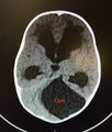
Obstructive hydrocephalus cause by a posterior fossa cyst in a 12 month old. Patient presented with loss of developmental milestones.

Management
Disposition
See Also
External Links
Sources
- ↑ Shprecher D. et al. Normal pressure hydrocephalus: diagnosis and treatment. Curr Neurol Neurosci Rep. 2008;8(5):371-376.