Ankle fracture: Difference between revisions
Mceledon83 (talk | contribs) |
Mceledon83 (talk | contribs) |
||
| Line 55: | Line 55: | ||
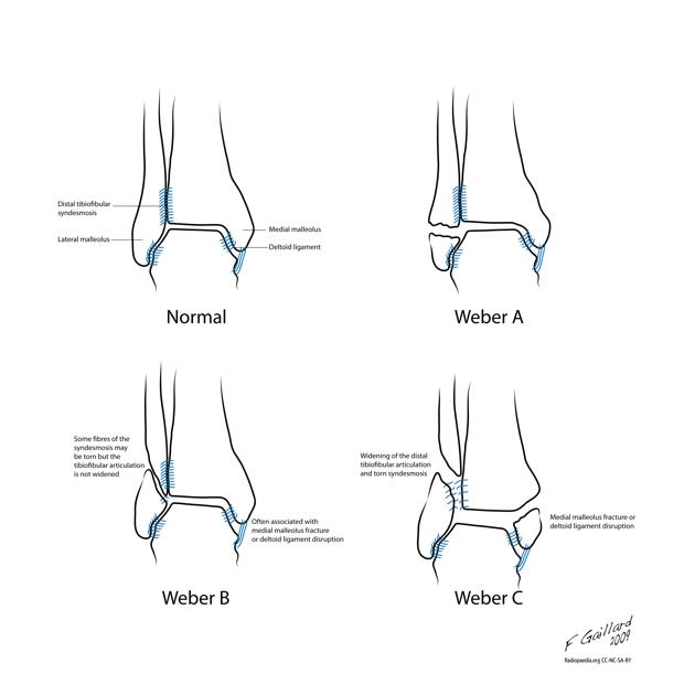
[[File:WeberclassRadioped.jpg|center|frame|300px]]<br> | [[File:WeberclassRadioped.jpg|center|frame|300px]]<br> | ||
==Management== | == Management == | ||
*Determined by stability of fx: | |||
**Stable, nondisplaced, isolated malleolar fx: Splint or cast, early wt bearing, RICE | *Determined by stability of fx: | ||
**Stable, nondisplaced, isolated malleolar fx: Splint or cast, early wt bearing, RICE | |||
**Unstable or displaced fx: Requires ORIF, ortho consult, reduce and splint | **Unstable or displaced fx: Requires ORIF, ortho consult, reduce and splint | ||
# Isolated lateral malleolar Fx | #Isolated lateral malleolar Fx | ||
## If stable (see Weber classification) treat like severe ankle sprain | ##If stable (see Weber classification) treat like severe ankle sprain | ||
## Signs of instability: | ##Signs of instability: | ||
###Displacement | ###Displacement >3mm | ||
###Associated medial malleolus fx | ###Associated medial malleolus fx | ||
###Signs of medial (deltoid) ligament disruption such as medial swelling, ecchymosis, or TTP | ###Signs of medial (deltoid) ligament disruption such as medial swelling, ecchymosis, or TTP | ||
###Widening of medial clear space (suggest deltoid ligament injury) | ###Widening of medial clear space (suggest deltoid ligament injury) | ||
# | #Isolated medial or posterior malleolar Fx | ||
## Must rule-out other injuries | ##Must rule-out other injuries | ||
## If non-displaced, isolated: | ##If non-displaced, isolated: | ||
### [[Short-Leg Posterior Splint]] (ankle at 90 | ###[[Short-Leg Posterior Splint]] (ankle at 90<sup>o</sup><sup></sup><sup></sup>) | ||
### Non-weight bearing | ###Non-weight bearing | ||
### Refer in 5-7d | ###Refer in 5-7d | ||
#Lateral malleolar fx with deltoid injury OR bimalleolar OR trimalleolar fx | #Lateral malleolar fx with deltoid injury OR bimalleolar OR trimalleolar fx | ||
##[[Short-Leg Posterior Splint]] (ankle at | ##[[Short-Leg Posterior Splint]] (ankle at 90<sup>o</sup>) | ||
##Immediate ortho consult in ED | ##Immediate ortho consult in ED | ||
Revision as of 00:18, 21 August 2013
Physical Exam
- Examine for ecchymoses, abrasions, or swelling
- Note skin integrity and areas of tenderness or crepitus over ankle
- Range joint passively and actively to evaluate for stability
- Examine Joints above and below the ankle
- Perform anterior drawer test (positive exam suggests torn ATFL)
- Always palpate entire length of fibula to rule-out Maisonneuve Fracture (fibulotibialis ligament tear)
- Perform a crossed-leg test to detect syndesmotic injury
- Evaluate integrity of Achilles tendon (Thompson test)
- Palpate midfoot and base of 5th metatarsal for tenderness
Diagnosis
- Ottawa Ankle Rules
- 3 views:
- AP
- Best for isolated lateral and medial malleolar fractures
- Oblique (mortise)
- Best for evaluating for unstable fracture or soft tissue injury
- At a point 1cm proximal to tibial plafond space between tib/fib should be ≤6cm
- Lateral
- Best for posterior malleolar fractures
- AP
- Determine if ankle fracture is:
- Unimalleolar
- Bimalleolar
- Trimalleolar
Classification (Danis-Weber System)
System based on level of the fibular fx and characterizes stability of fx
Type A (supination-adduction injury)
- Fibular Fx at or below level of ankle joint (talar mortise) without syndesmotic involvement
- Typically stable
- Deltoid ligament usually intact, medial malleolus usually fx
- A1: isolated
- A2: medial malleolus fx
- A3: posteromedial fx
Type B (supination-external rotation injury)
- Fibular Fx at level of ankle joint (talar mortise) w/ partial syndesmotic ligament injury
- Stability dictated by integrity of tibiofibular syndesmosis (no widening of distal tibiofibular articulation)
- Deltoid ligament may be torn, medial malleolus usually fx
- B1: isolated
- B2: medial lesion (either malleolus or ligament)
- B3: medial lesion and fx of posterolateral tibia
Type C (pronation-eversion injury)
- Fibular Fx above level of ankle joint (talar mortise) w/ complete syndesmotic disruption
- Unstable (widened distal tibiofibular articulation) and require surgical correction
- Deltoid ligament torn, medial malleolus fx
- C1: simple diaphyseal fibular fracture
- C2: complex diaphyseal fibular fracture
- C3: proximal fracture
Management
- Determined by stability of fx:
- Stable, nondisplaced, isolated malleolar fx: Splint or cast, early wt bearing, RICE
- Unstable or displaced fx: Requires ORIF, ortho consult, reduce and splint
- Isolated lateral malleolar Fx
- If stable (see Weber classification) treat like severe ankle sprain
- Signs of instability:
- Displacement >3mm
- Associated medial malleolus fx
- Signs of medial (deltoid) ligament disruption such as medial swelling, ecchymosis, or TTP
- Widening of medial clear space (suggest deltoid ligament injury)
- Isolated medial or posterior malleolar Fx
- Must rule-out other injuries
- If non-displaced, isolated:
- Short-Leg Posterior Splint (ankle at 90o)
- Non-weight bearing
- Refer in 5-7d
- Lateral malleolar fx with deltoid injury OR bimalleolar OR trimalleolar fx
- Short-Leg Posterior Splint (ankle at 90o)
- Immediate ortho consult in ED
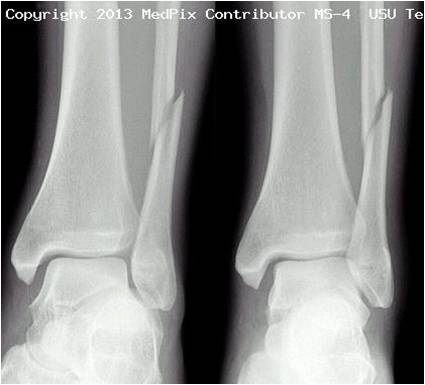
X-rays
See Also
- Ankle Sprain
- Ankle Fracture (Peds)
- [[Ottowa Ankle Rules]
- Maisonneuve Fracture
- Pilon Fracture
- Fracture (Main)
- Splinting
Source
- Tintinalli, Uptodate, Radiopaedia.org (Images by Dr. Frank Gaillard), Medpix Radiology Teaching Files (Images by Dr. Timothy Sanders)


