File:Computed tomograph of pulmonary vessels.jpg
Original file (1,413 × 1,067 pixels, file size: 288 KB, MIME type: image/jpeg)
Summary
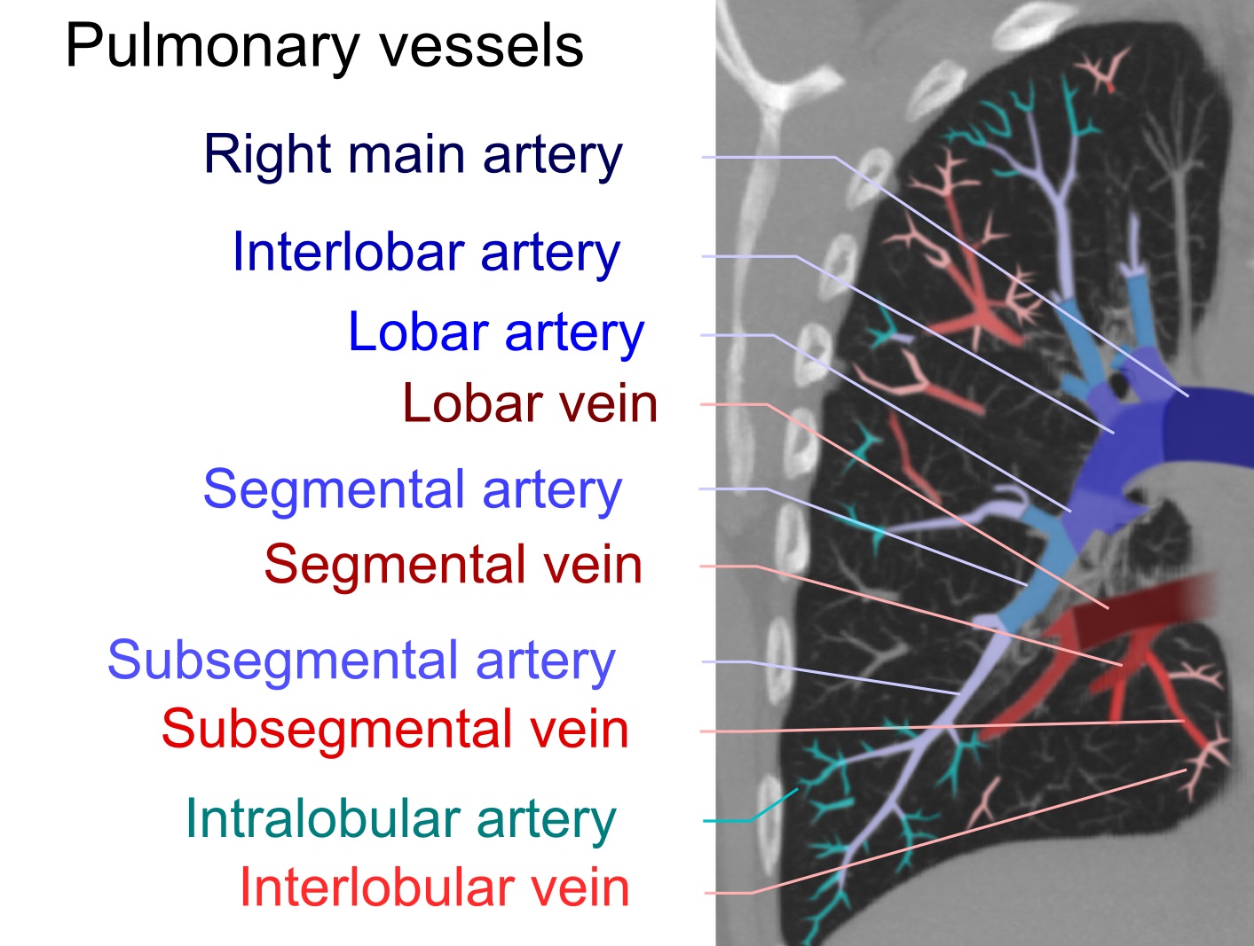
By Mikael Häggström - Own workeditReferences:Main pulmonary artery, segmental and subsegmental pulmonary arteries: Pulmonary Artery Anatomy. University of Virginia School of Medicine (2013). Retrieved on 2017-06-24.Right and left main pulmonary artery: Pulmonary Vasculature. University of Virginia School of Medicine (2013). Retrieved on 2017-06-24.Intralobular arteries: (2008). "Imaging of pulmonary emphysema: a pictorial review". Int J Chron Obstruct Pulmon Dis 3 (2): 193–204. PMID 18686729. PMC: 2629965.Interlobular veins: Page 58 in: David H. Trapnell (2016) Principles of X-Ray Diagnosis, Butterworth-Heinemann ISBN: 9781483195384., CC0, https://commons.wikimedia.org/w/index.php?curid=60990015
File history
Click on a date/time to view the file as it appeared at that time.
| Date/Time | Thumbnail | Dimensions | User | Comment | |
|---|---|---|---|---|---|
| current | 01:34, 8 January 2022 | | 1,413 × 1,067 (288 KB) | Rossdonaldson1 (talk | contribs) | By Mikael Häggström - Own workeditReferences:Main pulmonary artery, segmental and subsegmental pulmonary arteries: Pulmonary Artery Anatomy. University of Virginia School of Medicine (2013). Retrieved on 2017-06-24.Right and left main pulmonary arter... |
You cannot overwrite this file.
File usage
The following 3 pages use this file: